
관련상품
등록된 상품이 없습니다.
상품 정보
상품 상세설명
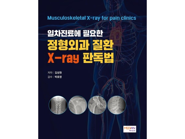
Contents
Ⅰ. CERVICAL
A. Anatomy of the Cervical
B. X-ray
1. AP view
a. Level check
b. Spondylosis
2. Lateral view
a. Alignment
b. Prevertebral soft tissue
c. Lordosis
d. Spondylosis
3. Extension/Flexion view
4. Oblique view
a. Foraminal stenosis
5. Open mouth view
C. Case
1. Infective spondylitis
2. Ossification posterior longitudinal ligament(OPLL)
3. Block vertebra
4. Ossification of nuchal ligament
Ⅱ. SHOULDER
A. Anatomy of the Shoulder
B. X-ray
1. AP view
a. Osteoarthritis
b. Acromioclavicular joint stress view
2. External/Internal rotation view
a. Hill-Sachs lesion
3. Grashey view(AP glenoid view)
a. Bankart lesion
4. Lateral scapula view(Y view)
a. Type of acromion
5. Axial view
6. Stryker notch view
C. Case
1. Rotator cuff disease에서의 X-ray 소견
2. Avascular necrosis(AVN
3. Scapular fracture
4. Osteoarthritis secondary to rheumatoid arthritis
5. Pseudocyst of the humerus
6. Osteosarcoma
7. Simple bone cyst
8. Pseudotumor deltoideus
9. Little leaguer’s shoulder
Ⅲ. ELBOW
A. Anatomy of the Elbow
B. X-ray
1. AP view
2. Lateral view
a. Radiocapitellar line
b. Anterior humeral line
c. Fat pad sign
3. Coyle’s view
4. Internal oblique view
5. External oblique view
C. Case
1. Osteoarthritis
2. Radius fracture
3. Calcific tendinitis
4. Loose body
5. Osteochondritis dissecans
6. Pediatric case
Ⅳ. WRIST
A. Anatomy of the Wrist
B. X-ray
1. PA view
a. Ulnar variance
b. Ulnar impaction syndrome
c. Kienbock disease
2. Lateral view
a. Pronator quadratus sign
3. Oblique view
4. Pisotriquetral view
5. Clenched fist view
6. Ulnar deviation view
a. Scaphoid fracture
7. Carpal tunnel view
a. Hook of the hamate fracture
C. Case
1. Rheumatoid arthritis
2. Finger osteoarthritis
3. Calcification
4. Avascular necrosis
5. Enchondroma
6. Fracture
7. Don’t touch lesion
Ⅴ. LUMBAR
A. Anatomy of the Lumbar
B. X-ray
1. AP view
a. Level check / Transitional vertebra
b. Scoliosis
c. Cobb’s angle
2. Lateral view
a. Disc herniation
b. Alignment
3. Extension/Flexion view
4. Oblique view
a. Facet joint osteoarthritis
b. Spondylolysis
c. Transverse process fracture
C. Case
1. Compression fracture
2. Spondylosis
3. Post op finding
4. Bertolotti syndrome
5. Infective spondylodiscitis
6. Schmorl node
7. Sacroiliac joint
7-1. Ankylosing spondylitis
7-2. Sacroiliac joint osteoarthritis
7-3. Osteitis condensans ilii
8. Don’t touch lesion
Ⅵ. HIP
A. Anatomy of the Hip
B. X-ray
1. AP view
a. Iliopectineal line, Shenton line, Ilioischial line
b. Fat pad
c. Lateral center-edge angle
d. Teardrop
e. Femoroacetabular impingement syndrome
2. Lateral view
C. Case
1. Osteoarthritis
2. Avascular necrosis (AVN)
3. Fracture
4. Post op finding
5. Pediatric case
6. Don’t touch lesion
Ⅶ. KNEE
A. Anatomy of the Knee
B. X-ray
1. AP view
2. Rosenberg view
3. Lateral view
a. Blumensaat line
b. Suprapatellar recess
c. Patella alta/baja
4. External rotation view
5. Internal rotation view
6. Skyline view
7. Posterior stress view
C. Case
1. Osteoarthritis
2. Loose body
3. ACL avulsion fracture
4. Osteochondritis dissecans
5. Subchondral insufficiency fracture of the knee
6. Osgood Schlatter’s disease
7. Osteochondroma
8. Don’t touch lesion
Ⅷ. ANKLE & FOOT
A. Anatomy of the Ankle & Foot
Ankle
B. X-ray
1. AP view
2. Mortise view
a. Clear space
3. Lateral view
a. Kager’s fat pad
b. Teardrop sign
c. Osteochondritis dissecans
4. Stress view(Anterior/Varus)
5. Axial calcaneus view
C. Case
1. Osteoarthritis(OA)
2. Anterior ankle impingement syndrome
3. Os trigonum syndrome
4. Stieda process
5. Fracture
6. Non-ossifying fibroma
Foot
B. X-ray
1. DP view
2. Lateral view
3. Oblique view
a. Lisfranc injury
b. Fracture
c. Apophysis
C. Case
1. Coalition
2. Osteoarthritis(OA)
3. Gout
4. Accessory navicular syndrome(= Prehallux syndrome)
5. Calcaneal spur
6. Hallux valgus
7. Pes planus(= Flat foot)
8. Haglund syndrome
9. Accessory ossicles
한페이지 지식
X-ray 기본 원리
Density, Echogenicity, Signal change
Ossification
Physis, Epiphysis, Apophysis
Calcification
X-ray 청구할 때 알아두어야 할 것
Osteopenia
Osteopenia-2
Spondyloarthritis
Apophysis
Fracture
Bursa vs Recess
Acute fracture vs Old fracture
Disuse osteopenia
사용후기
상품문의
배송정보
금액별 배송비
0원 이상~30,000원 미만 = 3,000원
30,000원 이상 = 0원